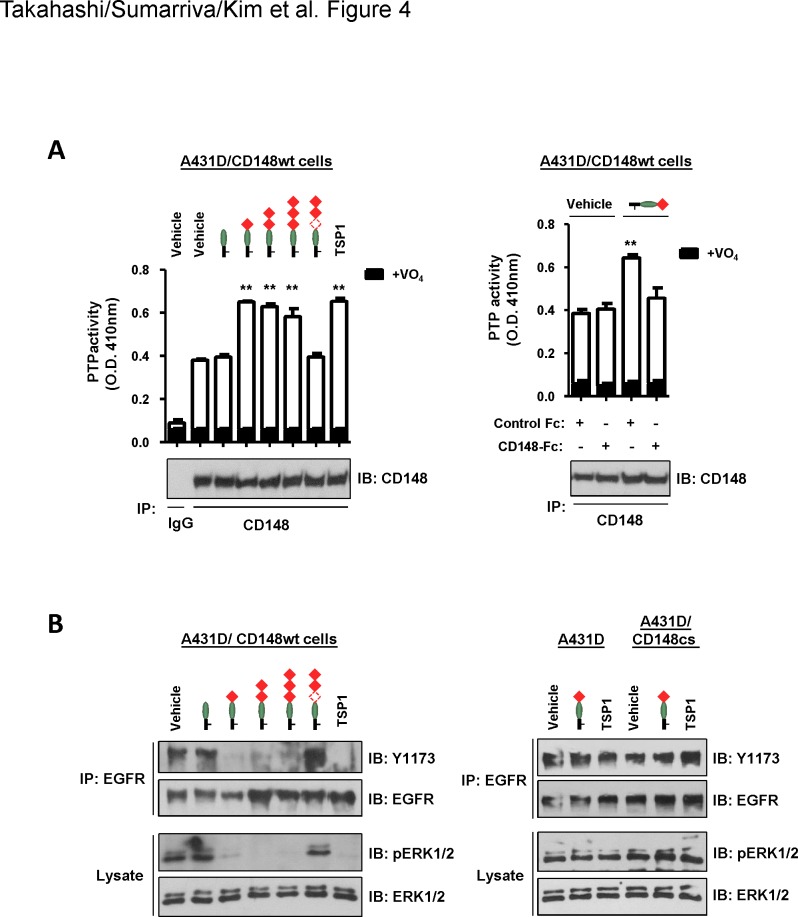
Fig 4. Trimeric TSP1 fragments that contain the 1st type 1 repeat increase CD148 catalytic activity and reduce tyrosine phosphorylation of EGFR and ERK1/2 in A431D/CD148wt cells.
(A) Left: A431D/CD148wt cells were treated with the indicated trimeric TSP1 fragments (12 nM) or whole TSP1 protein (12 nM) for 15 min. CD148 was immunoprecipitated using anti-CD148 antibody or class-matched control IgG. The washed immunocomplexes were subjected to a PTP activity assay with or without 1 mM sodium orthovanadate (VO4). The amount of CD148 in the immunocomplexes was evaluated by immunoblotting using anti-CD148 antibody (lower panel). The data show mean ± SEM of quadruplicate determinations. Representative data of five independent experiments is shown. ** P < 0.05 vs. vehicle-treated cells. Right: To assess the specificity of the effect, a trimeric TSP1 fragment containing the procollagen domain and the 1st type 1 repeat was added to A431D/CD148wt cells with 11.3 nM of CD148-Fc or control Fc (Fc alone), then CD148 catalytic activity was assessed as in left panel. The data show mean ± SEM of quadruplicate determinations. Representative data of five independent experiments is shown. ** P < 0.05 vs. vehicle-treated cells. Note: CD148-Fc, but not control Fc, abolishes the activity of the TSP1 fragment to increase CD148 catalytic activity. (B) Left: A431D/CD148wt cells were treated with the indicated trimeric TSP1 fragments (12 nM) or whole TSP1 protein (12 nM) for 15 min. Tyrosine phosphorylation of EGFR (immunoprecipitated) and ERK1/2 was assessed by immunoblotting using the phopho-specific EGFR (Y1173) or ERK1/2 (T202/Y204) antibodies. The membranes were reprobed with antibodies to total EGFR or ERK1/2. Representative data of four independent experiments is shown. Right: A431D and A431D/CD148cs cells were treated with a trimeric TSP1 fragment (12 nM) that contains the procollagen domain and the 1st type 1 repeat or whole TSP1 protein (12 nM), and tyrosine phosphorylation of EGFR (immunoprecipitated) and ERK1/2 was assessed as in left panel. Representative data of four independent experiments is shown. Note: No effects are observed in A431D and A431D/CD148cs cells.

