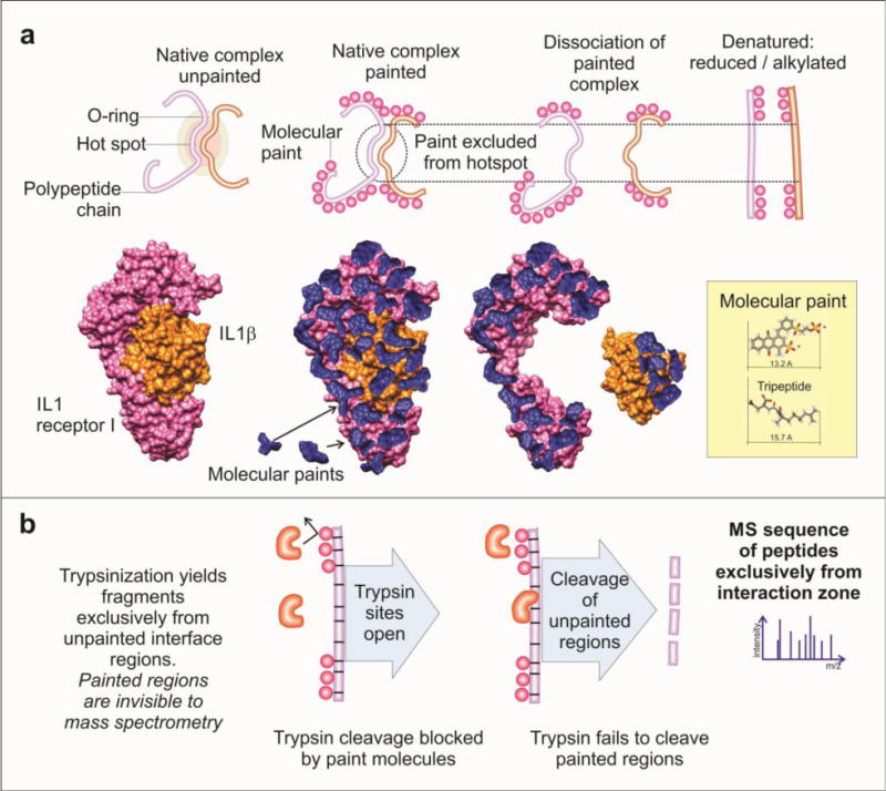
Figure 5. Protein painting.

a) Protein–protein complexes are painted using surface binding molecular paints. The area of protein–protein interaction is not accessible to the solvent, so these regions remain unpainted and are available for trypsin digestion following dissociation and denaturation of the complex. A model of the IL1β complex is illustrated before and after undergoing protein painting. b) The molecular paints bind to the regions that are solvent-accessible and block trypsin cleavage sites. After digestion, the resulting peptides will be derived exclusively from the region of protein–protein interaction and can be further processed and identified by mass spectrometry [21]. Modified from Luchini et al [25].
