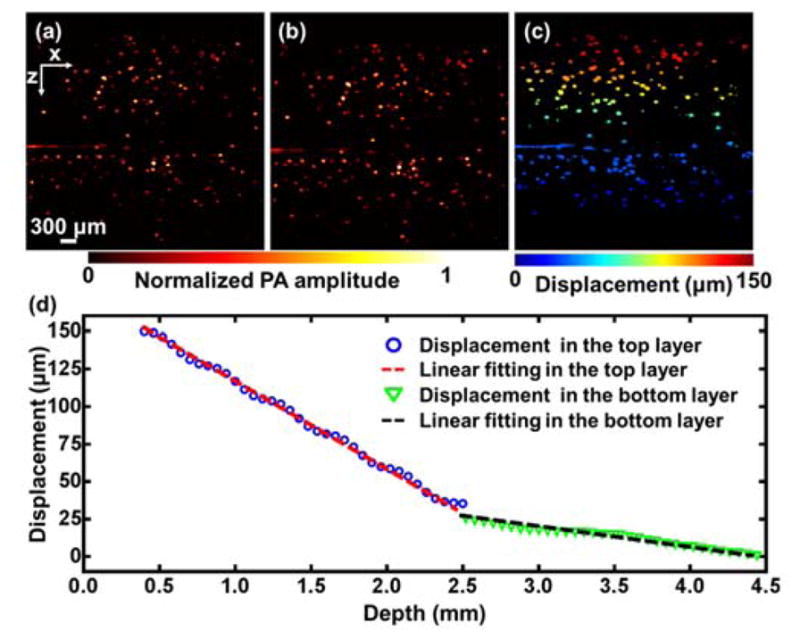
Fig. 3.

Strain measurement of a bilayer gelatin phantom by photoacoustic elastography. (a–b) PA images of a bilayer gelatin phantom mixed with 50 μm microspheres acquired (a) before and (b) after compression. (c) Displacement image obtained from (a) and (b). (d) Average displacement versus depth. The data was fitted by a linear function for each layer.
