Fig. 5.

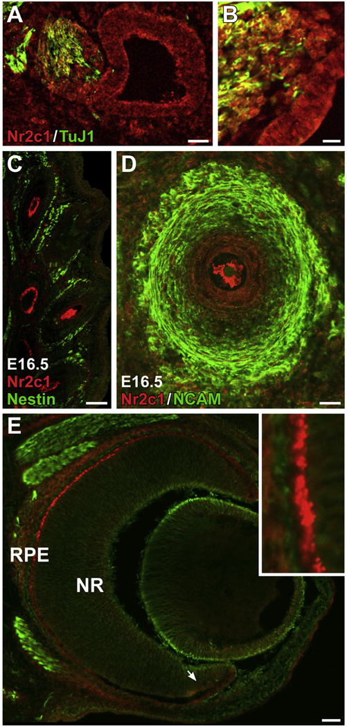
A–B. Nr2c1 is expressed in the placodally-derived neurepithelium of the otic vesicle at E10.5, as well as in the adjacent otic ganglion, as viewed in a saggital section. B. Higher magnification view of otic ganglia/otic vesicle from adjacent section. C–D. Expression of Nr2c1 in support cells of the facial vibrissae, as observed in a coronal section of an E16.5 embryo. C. Low-magnification image across multiple vibrissae shows that Nr2c1 labeled cells have distinctive morphologies, depending on the depth of the cross-section of the vibrissae. D. Confocal optical section (20×) illustrating support cells. E. Nr2c1 is robustly expressed in the presumptive retinal pigment epithelia of the eye at E14.5. Arrow: ciliary marginal zone. Inset: 5× magnification.
