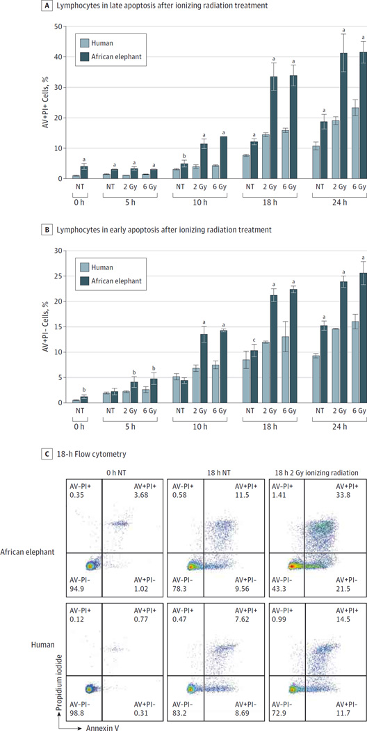
Figure 3. African Elephant and Human Peripheral Blood Lymphocytes and Sensitivity to Ionizing Radiation.
A, The percentage of late apoptosis (annexin V positive [AV+] and propidium iodide positive [PI+]) and B, early apoptosis (AV+PI−) in elephant peripheral blood lymphocytes compared with human peripheral blood lymphocytes in response to 2 Gy and 6 Gy of ionizing radiation are graphed. Significant differences computed with a 2-sided t test between human and elephant at 0, 5, 10, 18, and 24 hours are indicated. Error bars represent 95% CIs. C, Representative scatter plots from flow cytometry are shown from the 0- and 18-hour time points. NT indicates no treatment.
aP <. 001.
bPanel A: NT at 10 hours, P = .008. Panel B: NT at 0 hours, P = .002; 2 Gy at 5 hours, P = .003; 6 Gy at 5 hours, P = .004.
cP = .03.

