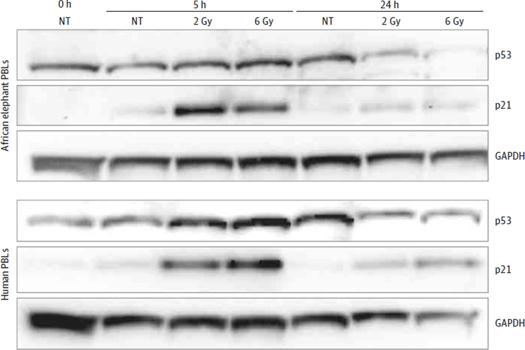
Figure 6. p21 and p53 Protein Expression After Ionizing Radiation.
Western blot at the indicated time points after ionizing radiation shows p21 and p53 protein expression in elephant and human lymphocytes. The p53 antibody detects only nonphosphorylated protein. GAPDH indicates glyceraldehyde 3-phosphate dehydrogenase, a protein-loading control; PBL, peripheral blood lymphocyte; NT, no treatment.

