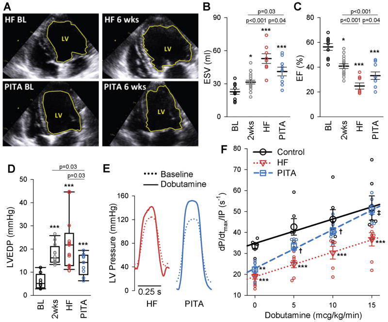
Figure 1. Pacemaker induced transient asynchrony (PITA) improves in vivo cardiac function.
(A) Example end-diastolic echocardiographic images at baseline (BL) and after 6-weeks (end of study) of dog hearts in the heart failure (HF) and PITA treated group. The left-ventricle (LV) is outlined in yellow. (B and C) LV end-systolic volume (ESV) (B) and ejection fraction (EF) (C) assessed by echocardiography at baseline (BL, n = 10), after two weeks of atrial pacing (n = 18), at the end of the six-week atrial pacing protocol (HF, n = 8), and at the end of six-week PITA protocol (n = 9). (D) LV end-diastolic pressure (LVEDP) from hemodynamic studies (n: Con = 8, 2wks = 12, HF = 13, PITA = 10 dogs). EDP data were non-normal, and are displayed as box plots. Data were log10-transformed before one-way ANOVA. Direct comparison to 2wks group made by t-test). (E) Example LV pressure waveforms in HF and PITA hearts at baseline and with 15 mcg/kg/min dobutamine. (F) Dobutamine dose-effect on LV contractility (dP/dtmax/IP) (n: Control = 6, HF = 9, PITA = 8 dogs, some doses missing for individual dogs). Data points indicate individual animals at each dose, symbols are means ± SEM. *p<0.05, **p<0.01, ***p<0.001 vs. Control; †p<0.05, ‡p<0.001 vs. HF by one-way ANOVA and Holm Sidak post-hoc test.

