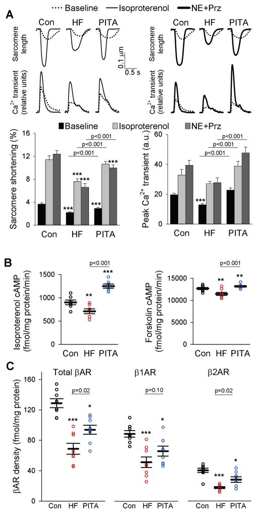
Figure 2. Myocyte function is depressed in HF after β-adrenergic receptor stimulation, but near normal with PITA.
(A) Example tracings of sarcomere shortening and intracellular calcium transients from LV lateral wall myocytes isolated from healthy control, HF, and PITA dogs at baseline and after isoproterenol stimulation (0.1 μM) or after norepinephrine and prazosin stimulation (NE, 0.1 μM; Prz, 1 μM). Sarcomere shortening and intracellular calcium are quantified below as means ± SEM (n = 4–7 dogs in each group, cells/dog: 5.8 ± 0.4, mean ± SEM). For all three groups, Iso and NE+Prz sarcomere shortening and peak Ca2+ transient data are p<0.05 versus respective baseline; ***p<0.001 vs. Control by two-way ANOVA and Holm-Sidak post-hoc test. (B) Cyclic AMP activity after isoproterenol or forskolin (FSK) stimulation. Data are individual dogs (n = 8), with means ± S.E.M. (C) Plasma membrane β-AR, β1-AR, and β2-AR density. Data are individual dogs and means ± SEM (n = 8 per group). In B and C, *p<0.05, **p<0.01, ***p<0.001 vs. Control by one-way ANOVA and Holm-Sidak post-hoc test.

