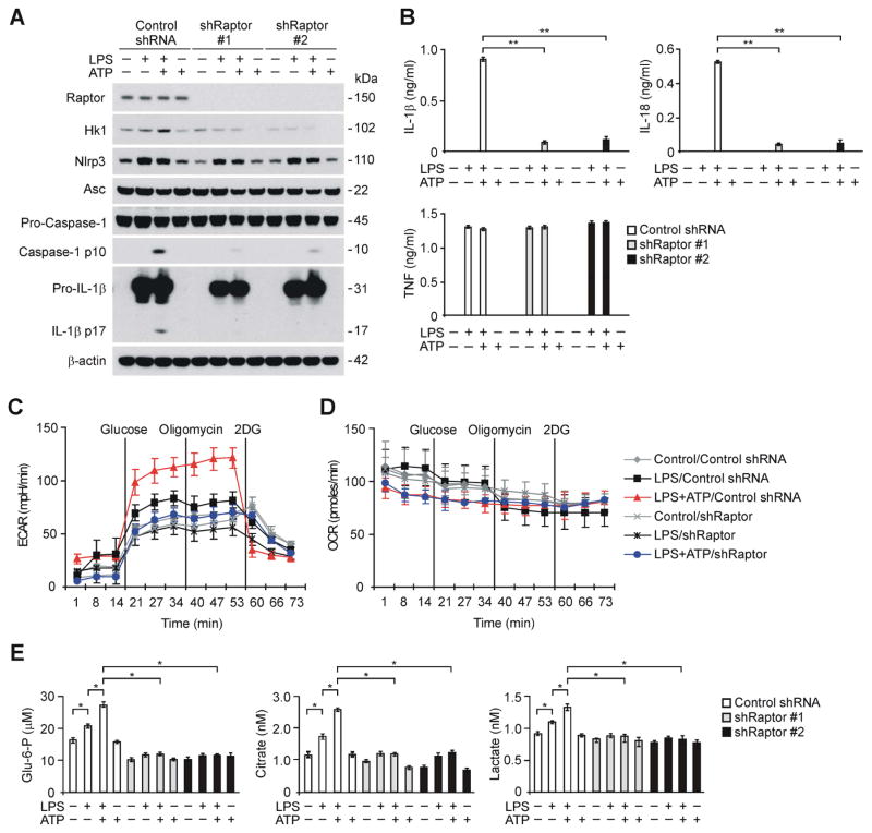
Figure 3. Deficiency of Raptor/mTORC1 suppresses caspase-1 activation, HK1 expression and glycolysis during NLRP3 inflammasome activation.
(A) Immunoblot analysis for Raptor, HK1, caspase-1 and IL-1β in cell lysates from wild type mouse peritoneal macrophages transduced with lentiviruses expressing non-target shRNA (Control shRNA) or two independent shRNA for Raptor (shRaptor #1 and #2), and stimulated with LPS and ATP. β-actin served as the standard. (B) ELISA assay for IL-1β, IL-18 and TNF secretion from A. **P<0.01 by ANOVA. (C) ECAR was measured in wild type peritoneal macrophages transduced with lentiviruses expressing non-target shRNA (Control shRNA) or shRNA for Raptor (shRaptor), and stimulated with LPS and ATP. Data are mean ± s.d. (D) OCR was measured in wild type peritoneal macrophages transduced with lentiviruses expressing non-target shRNA (Control shRNA) or shRNA for Raptor (shRaptor), and stimulated with LPS and ATP. Data are mean ± s.d. (E) Glucose-6-phosphate, citrate and lactate production assay from wild type peritoneal macrophages transduced with lentiviruses expressing non-target shRNA (Control shRNA) or two independent shRNA for Raptor (shRaptor #1 and #2), and stimulated with LPS and ATP. *P<0.05 by ANOVA. See also Figure S3.

