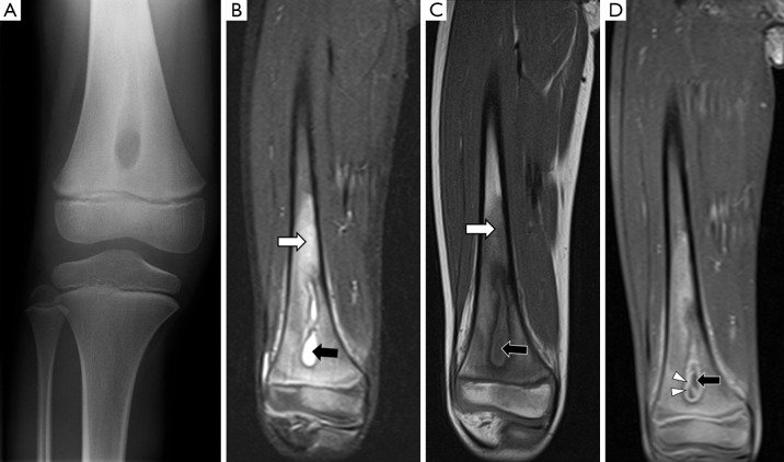
Figure 3.
A 6-year-old girl with no history of trauma presents with pain and swelling in her right knee. (A) The anteroposterior radiograph shows a well-circumscribed lucent lesion with sclerotic margins in the right distal femur metaphysis, suspicious for an intraosseous abscess; (B) coronal STIR image of the right femur shows that the lesion is within the medullary cavity and has high signal (black arrow). The bone marrow of the distal diaphysis and metaphysis has diffuse high signal (white arrow) compared to the mid-diaphysis, representing bone marrow oedema; (C) coronal T1W image shows that the intraosseous lesion has central heterogeneous low signal (black arrow). The bone marrow of the distal diaphysis and metaphysis has diffuse low signal consistent with oedema (white arrow). Note the distinct margin between normal and abnormal marrow, suggestive of bone marrow oedema due to osteomyelitis rather than reactive osteitis; (D) coronal fat-suppressed T1W image after administration of intravenous contrast shows that the lesion has central low signal (black arrow) and peripheral enhancement (white arrowheads). The central low signal represents pus and the peripherally enhancing areas represent hypervascular granulation tissue. This confirms an intraosseous abscess. Its location in the metaphysis is characteristic for haematogenous osteomyelitis.

