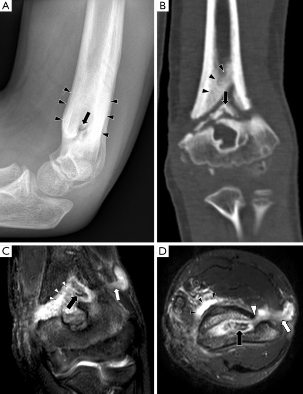
Figure 4.
Chronic osteomyelitis in a 9-year-old boy with a non-united left distal humerus fracture. (A) The lateral radiograph shows marked periosteal thickening (black arrowheads) and a central sclerotic lesion with a lucent rim (black arrow); (B) coronal CT with bone windows shows a sclerotic fragment of bone which is separate from the rest of the humerus (black arrow), consistent with a sequestrum. Cortical thickening is also noted (black arrowheads); this represents an involucrum which is a result of periosteal new bone formation. These findings were not present on initial images taken at the time of the fracture; (C) coronal STIR image shows the low signal sequestrum (black arrow) surrounded by high signal pus and granulation tissue (white arrowheads). There is a sinus tract draining pus to the skin surface (white arrow); (D) axial fat-suppressed T2 image demonstrates that the pus surrounding the sequestrum (black arrow) communicates with the sinus tract (white arrow) via a cloaca (white arrowhead). There is also a soft tissue fluid collection anteromedial to the humerus (black arrowheads). (Images courtesy of Dr. Asif Saifuddin, Royal National Orthopaedic Hospital, Stanmore).

