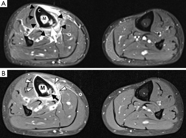
Figure 5.
Osteomyelitis in the right tibia of a 44-year-old male. (A) Axial STIR image of both legs. There is high signal in the medullary cavity of the right tibia (M) compared to the normal low signal medulla of the left tibia. This could represent either bone marrow oedema or an intramedullary abscess. There is also circumferential periosteal elevation and high signal (black arrowheads), suggesting a periosteal reaction with a possible subperiosteal abscess. Within the tibial cortex (C), there is a focal area of high signal intensity (white arrow), suspicious for an intraosseous abscess; (B) axial fat-suppressed T1W image following intravenous contrast. The cortical lesion (black arrow) has central low signal and peripheral enhancement, confirming the suspicion of a cortical abscess. There is uniform enhancement of the bone marrow (M) and periosteum (white arrowheads), consistent with bone marrow oedema and a periosteal reaction. The absence of central low signal in these regions excludes intramedullary and subperiosteal abscesses.

