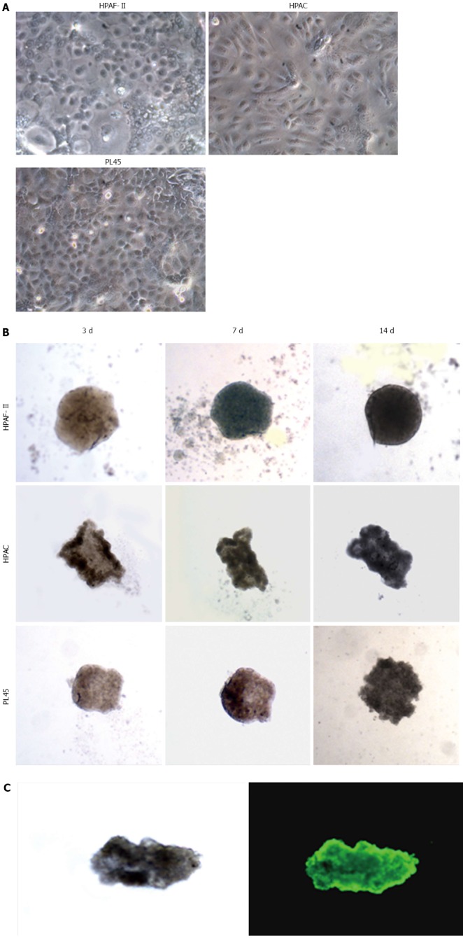
Figure 1.

Morphology of pancreatic adenocarcinoma cells grown in 2D-monolayers and 3D-spheroids. A: Micrograph from inverted microscope showing the epithelial morphology of HPAF-II, HPAC, and PL45 cells grown in 2D-monolayers. Original magnification: 20 ×; B: 3D-spheroids observed under inverted microscope after 3, 7, and 14 d. HPAF-II spheroids were more rounded and uniformly dense; by contrast, HPAC and PL45 spheroids displayed an irregular shape. Original magnification: 10 ×; C: Representative 3D-spheroid under inverted microscope and after incubation with calcein-AM. Original magnification: 10 ×.
