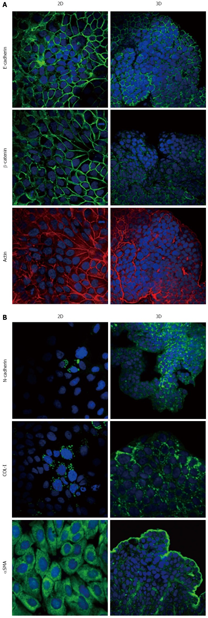
Figure 6.

Expression of epithelial-to-mesenchymal transition-related markers in HPAC cells. Micrographs using a confocal microscope showing epithelial (A) and mesenchymal markers (B) in HPAC cells grown in 2D-monolayers and 3D-spheroids. Original magnification: 60 ×.
