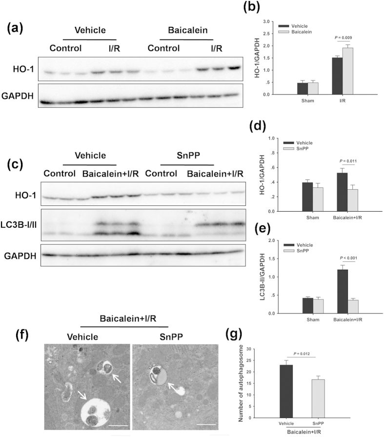
Figure 4. HO-1 mediates baicalein-induced autophagy.
Rats were pretreated with SnPP (50 mg/kg, IP) 0.5 h prior to baicalein treatment (100 mg/kg, IP) and killed at 6 h after reperfusion. (a) HO-1 protein expression in the ischemic lobes was examined by western blot analysis. (b) Densitometric analysis of HO-1 expression. (c) The expression levels of HO-1 and LC3B in the ischemic lobes were examined by western blot analysis. (d) Densitometric analysis of HO-1 expression. (e) Densitometric analysis of LC3B-II expression. (f) Representative transmission electron micrographs showing autophagosomes in the ischemic lobes at 6 h of reperfusion. Autophagosomes are indicated by arrows. Scale bars 1 µm. (g) Quantification of autophagosomes. The data are shown as the mean ± SD. n = 6 per group.

