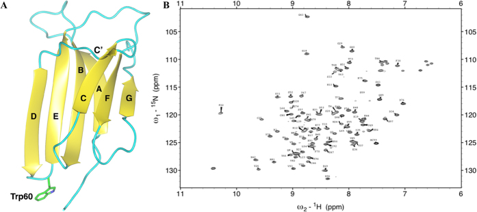
Figure 1. NMR assigment of β2m.
(A) Ribbon representation of the crystal structure of human β 2m (pdb code 2YXF) with β -strands labeled according to standard nomenclature. W60 is shown as sticks. (B) HSQC [1H, 15N] spectrum recorded at 11.7 T (500.13 MHz for 1H), 310K, of [U-13C, U-15N W60G β 2m] 0.5 mM dissolved in 70 mM phosphate buffer at pH = 6.6 and 100 mM NaCl.

