Abstract
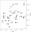
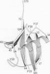
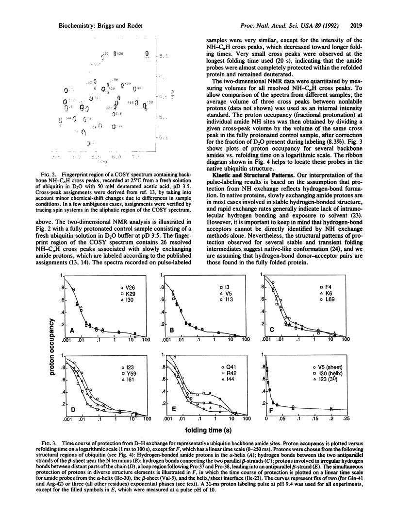
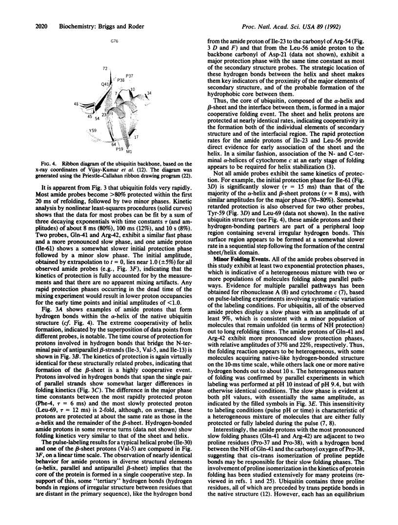
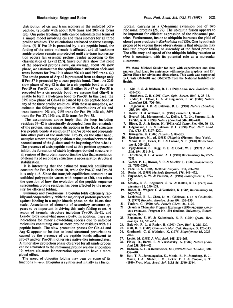
The formation of hydrogen-bonded structure in the folding reaction of ubiquitin, a small cytoplasmic protein with an extended beta-sheet and an alpha-helix surrounding a pronounced hydrophobic core, has been investigated by hydrogen-deuterium exchange labeling in conjunction with rapid mixing methods and two-dimensional NMR analysis. The time course of protection from exchange has been measured for 26 back-bone amide protons that form stable hydrogen bonds upon refolding and exchange slowly under native conditions. Amide protons in the beta-sheet and the alpha-helix, as well as protons involved in hydrogen bonds at the helix/sheet interface, become 80% protected in an initial 8-ms folding phase, indicating that the two elements of secondary structure form and associate in a common cooperative folding event. Somewhat slower protection rates for residues 59, 61, and 69 provide evidence for the subsequent stabilization of a surface loop. Most probes also exhibit two minor phases with time constants of about 100 ms and 10 s. Only two of the observed residues, Gln-41 and Arg-42, display significant slow folding phases, with amplitudes of 37% and 22%, respectively, which can be attributed to native-like folding intermediates containing cis peptide bonds for Pro-37 and/or Pro-38. Compared with other proteins studied by pulse labeling, including cytochrome c, ribonuclease, and barnase, the initial formation of hydrogen-bonded structure in ubiquitin occurs at a more rapid rate and slow-folding species are less prominent.
Full text
PDF
Images in this article
Selected References
These references are in PubMed. This may not be the complete list of references from this article.
- Baldwin R. L., Roder H. Characterizing protein folding intermediates. Curr Biol. 1991 Aug;1(4):218–220. doi: 10.1016/0960-9822(91)90061-z. [DOI] [PubMed] [Google Scholar]
- Butt T. R., Jonnalagadda S., Monia B. P., Sternberg E. J., Marsh J. A., Stadel J. M., Ecker D. J., Crooke S. T. Ubiquitin fusion augments the yield of cloned gene products in Escherichia coli. Proc Natl Acad Sci U S A. 1989 Apr;86(8):2540–2544. doi: 10.1073/pnas.86.8.2540. [DOI] [PMC free article] [PubMed] [Google Scholar]
- Bycroft M., Matouschek A., Kellis J. T., Jr, Serrano L., Fersht A. R. Detection and characterization of a folding intermediate in barnase by NMR. Nature. 1990 Aug 2;346(6283):488–490. doi: 10.1038/346488a0. [DOI] [PubMed] [Google Scholar]
- Di Stefano D. L., Wand A. J. Two-dimensional 1H NMR study of human ubiquitin: a main chain directed assignment and structure analysis. Biochemistry. 1987 Nov 17;26(23):7272–7281. doi: 10.1021/bi00397a012. [DOI] [PubMed] [Google Scholar]
- Englander S. W., Kallenbach N. R. Hydrogen exchange and structural dynamics of proteins and nucleic acids. Q Rev Biophys. 1983 Nov;16(4):521–655. doi: 10.1017/s0033583500005217. [DOI] [PubMed] [Google Scholar]
- Finley D., Bartel B., Varshavsky A. The tails of ubiquitin precursors are ribosomal proteins whose fusion to ubiquitin facilitates ribosome biogenesis. Nature. 1989 Mar 30;338(6214):394–401. doi: 10.1038/338394a0. [DOI] [PubMed] [Google Scholar]
- Grathwohl C., Wüthrich K. The X-Pro peptide bond as an nmr probe for conformational studies of flexible linear peptides. Biopolymers. 1976 Oct;15(10):2025–2041. doi: 10.1002/bip.1976.360151012. [DOI] [PubMed] [Google Scholar]
- Kim P. S., Baldwin R. L. Intermediates in the folding reactions of small proteins. Annu Rev Biochem. 1990;59:631–660. doi: 10.1146/annurev.bi.59.070190.003215. [DOI] [PubMed] [Google Scholar]
- Kuwajima K. The molten globule state as a clue for understanding the folding and cooperativity of globular-protein structure. Proteins. 1989;6(2):87–103. doi: 10.1002/prot.340060202. [DOI] [PubMed] [Google Scholar]
- Lenkinski R. E., Chen D. M., Glickson J. D., Goldstein G. Nuclear magnetic resonance studies of the denaturation of ubiquitin. Biochim Biophys Acta. 1977 Sep 27;494(1):126–130. doi: 10.1016/0005-2795(77)90140-4. [DOI] [PubMed] [Google Scholar]
- Levitt M. Effect of proline residues on protein folding. J Mol Biol. 1981 Jan 5;145(1):251–263. doi: 10.1016/0022-2836(81)90342-9. [DOI] [PubMed] [Google Scholar]
- Molday R. S., Englander S. W., Kallen R. G. Primary structure effects on peptide group hydrogen exchange. Biochemistry. 1972 Jan 18;11(2):150–158. doi: 10.1021/bi00752a003. [DOI] [PubMed] [Google Scholar]
- Pace C. N. Determination and analysis of urea and guanidine hydrochloride denaturation curves. Methods Enzymol. 1986;131:266–280. doi: 10.1016/0076-6879(86)31045-0. [DOI] [PubMed] [Google Scholar]
- Redman K. L., Rechsteiner M. Identification of the long ubiquitin extension as ribosomal protein S27a. Nature. 1989 Mar 30;338(6214):438–440. doi: 10.1038/338438a0. [DOI] [PubMed] [Google Scholar]
- Roder H., Elöve G. A., Englander S. W. Structural characterization of folding intermediates in cytochrome c by H-exchange labelling and proton NMR. Nature. 1988 Oct 20;335(6192):700–704. doi: 10.1038/335700a0. [DOI] [PMC free article] [PubMed] [Google Scholar]
- Roder H. Structural characterization of protein folding intermediates by proton magnetic resonance and hydrogen exchange. Methods Enzymol. 1989;176:446–473. doi: 10.1016/0076-6879(89)76024-9. [DOI] [PubMed] [Google Scholar]
- Roder H., Wagner G., Wüthrich K. Individual amide proton exchange rates in thermally unfolded basic pancreatic trypsin inhibitor. Biochemistry. 1985 Dec 3;24(25):7407–7411. doi: 10.1021/bi00346a056. [DOI] [PubMed] [Google Scholar]
- Roder H., Wüthrich K. Protein folding kinetics by combined use of rapid mixing techniques and NMR observation of individual amide protons. Proteins. 1986 Sep;1(1):34–42. doi: 10.1002/prot.340010107. [DOI] [PubMed] [Google Scholar]
- Tanford C. Protein denaturation. C. Theoretical models for the mechanism of denaturation. Adv Protein Chem. 1970;24:1–95. [PubMed] [Google Scholar]
- Udgaonkar J. B., Baldwin R. L. Early folding intermediate of ribonuclease A. Proc Natl Acad Sci U S A. 1990 Nov;87(21):8197–8201. doi: 10.1073/pnas.87.21.8197. [DOI] [PMC free article] [PubMed] [Google Scholar]
- Udgaonkar J. B., Baldwin R. L. NMR evidence for an early framework intermediate on the folding pathway of ribonuclease A. Nature. 1988 Oct 20;335(6192):694–699. doi: 10.1038/335694a0. [DOI] [PubMed] [Google Scholar]
- Vijay-Kumar S., Bugg C. E., Cook W. J. Structure of ubiquitin refined at 1.8 A resolution. J Mol Biol. 1987 Apr 5;194(3):531–544. doi: 10.1016/0022-2836(87)90679-6. [DOI] [PubMed] [Google Scholar]
- Weber P. L., Brown S. C., Mueller L. Sequential 1H NMR assignments and secondary structure identification of human ubiquitin. Biochemistry. 1987 Nov 17;26(23):7282–7290. doi: 10.1021/bi00397a013. [DOI] [PubMed] [Google Scholar]


