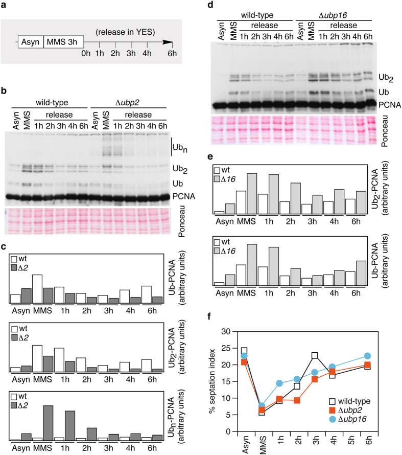
Figure 2. Fission yeast cells lacking ubp2+ or ubp16+ deubiquitylate ubPCNA after MMS-induced DNA damage.
(a) Experimental design; exponentially growing cultures of wild-type, Δ ubp2, and Δ ubp16 strains were treated with 0.02% MMS and then released. Samples were taken at indicated intervals for cell cycle and Western analysis. (b) Western blot analysis in wild-type and Δ ubp2 mutant cells. ubPCNA signals were quantified and normalized to loading controls. (c) Quantification is shown in bar diagrams. (d) Western blot analysis in wild-type and Δ ubp16 mutant cells. As was previously performed, ubPCNA signals were quantified and normalized to loading controls. (e) Quantification is shown in bar diagrams. (f) Plots of septated (septation index) cells of the indicated strains in (b,d) are shown.

