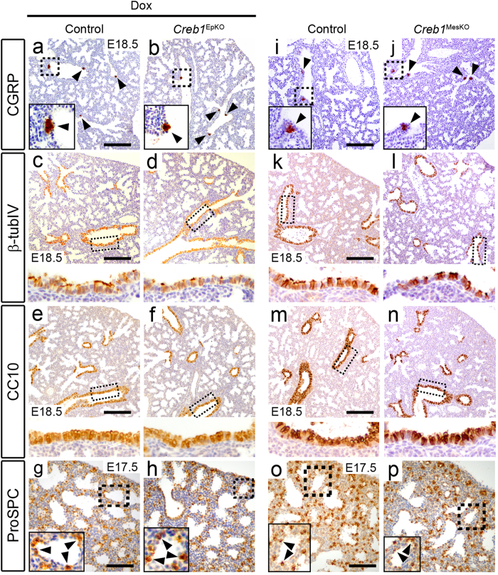
Figure 3. Normal epithelial differentiation in Creb1EpKO lungs.
Immunohistochemistry for the proximal epithelial markers CGRP (a,b, i,j), β-tubulin IV (c,d,k,l), and CC10 (e,f,m,n) in E18.5 Creb1EpKO and Creb1MesKO lungs. Immunohistochemistry for the distal epithelial (type-II AEC) marker ProSPC (g,h,o,p) in E17.5 Creb1EpKO and E18.5 Creb1MesKO lungs. No differences in expression or localization was observed for proximal or epithelial markers. Boxed areas are magnified either in insets (CGRP, ProSPC) or below the main image (β-tubulin IV, CC10), while arrowheads indicate marker-positive cells. All images are representative of at least three animals per genotype. Scale bars: a-f, i-n; 180 μm. g,h,o,p; 90 μm.

