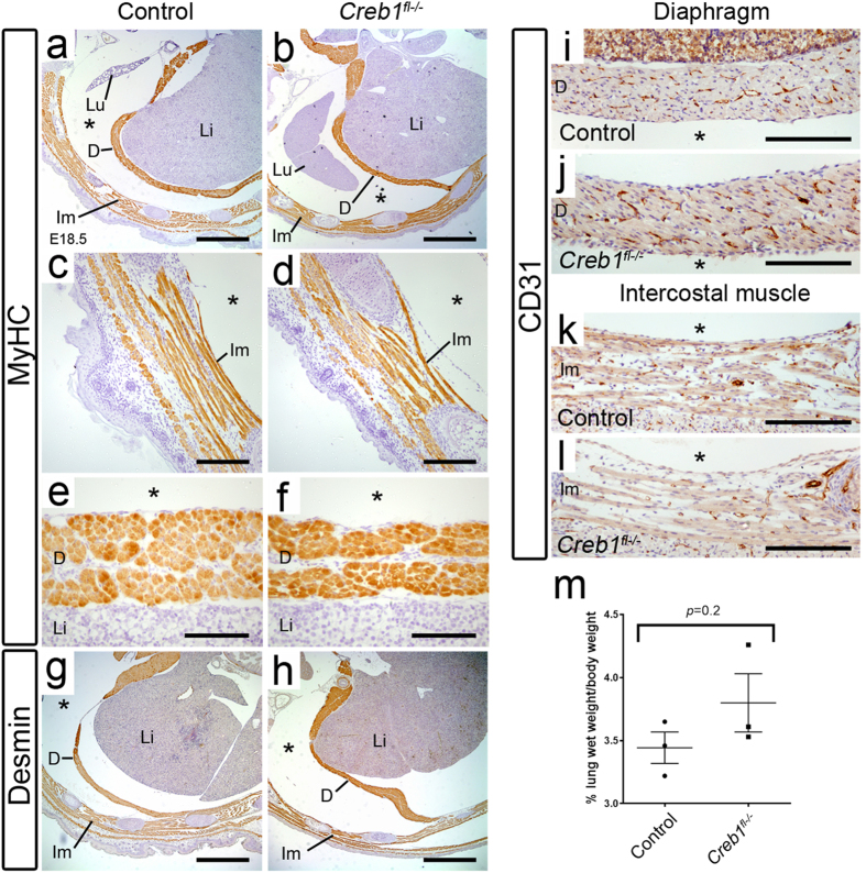
Figure 6. Diaphragm and intercostal musculature are normally developed in Creb1fl−/− fetal mice.
Immunohistochemistry for the muscle markers MyHC (a–f), desmin (g,h) and CD31 (i–l) in transverse sections through thoracic vertebrae in E18.5 control and Creb1fl−/− fetal mice. Magnified images of MyHC-positive intercostal (c,d) and diaphragm (e,f) musculature. Intercostal and diaphragm musculature appear normally developed in Creb1fl−/− fetal mice. (m) Scatterplot showing the percentage fetal lung wet weight/body weight in E18.5 control and Creb1fl−/− littermates (n = 3). Error bars represent SEM. All images are representative of at least three animals per genotype. Scale bars: a,b,g,h; 900 μm, c,d,i-l; 180 μm, e,f; 90 μm. Lu: lung, Li: liver, D: diaphragm, Im: intercostal muscle, “*”indicates thoracic cavity.

