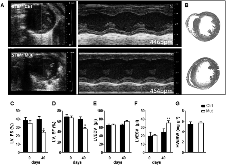
Figure 1. Echocardiography at 0 and 40 days after tamoxifen injection reveals decreased cardiac performances in STIM1 mutant mice.
(A) Representative ultrasound images of LV (short-axis) in B-mode and M-mode (at the level of the papillary muscles) from STIM1 mutant and control mice. (B) Representative heart cross-sections from control mice (upper panel) and STIM1 mutant mice (lower panel). (C–F) Averaged LV parameters obtained from analysis of echocardiograms: (C) fractional shortening (FS); (D) ejection fraction (EF); (E) left ventricular end-diastolic volume (LV-EDV); (F) left ventricular end-systolic volume (LV-ESV). (G) Ventricular weight/body weight ratio (mg of left ventricle/g of body weight), data taken 40 days post injection, (control hearts n = 6; STIM1 mutant, n = 5). Values represent mean ± SEM, echo data were obtained from n = 11 controls and n = 16 STIM1 mutant mice. *P < 0.05, **P < 0.01.

