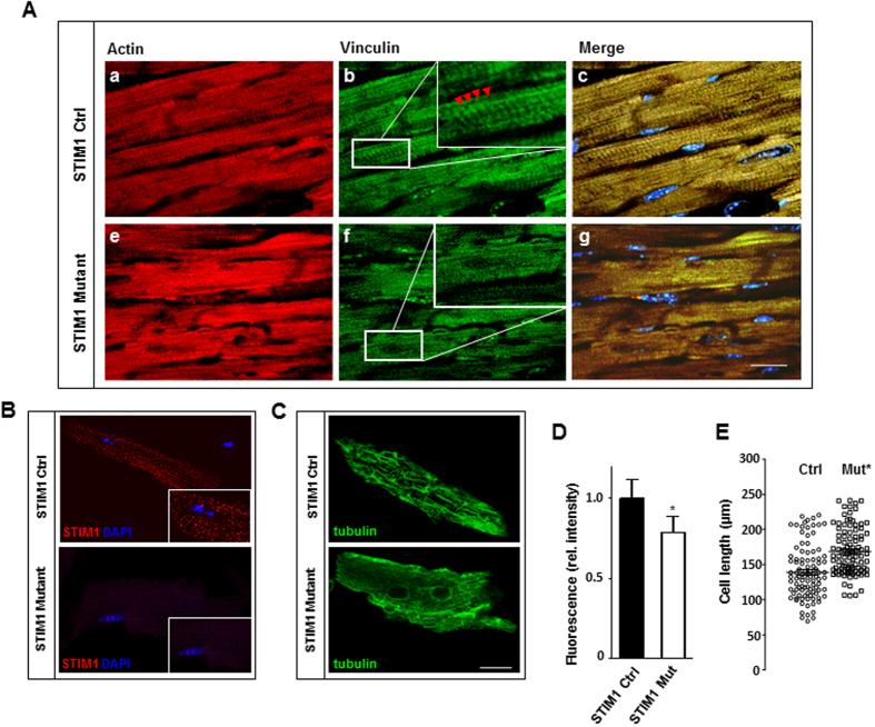
Figure 3. STIM1 genetic deletion results in heterogeneous vinculin distribution and myofibrillar rearrangements.
Hearts were harvested 40 days after tamoxifen treatment. (A) Representative immunostaining of heart sections. Actin was labeled with rhodamine-phalloidin (red) to identify cardiomyocytes (a–e), vinculin, in green, is detected by anti-vinculin antibody (b–f), nuclei in blue are stained with DAPI (c–g). In control hearts vinculin immunofluorescence staining shows costameres with intensely stained intercalated disks indicated by red arrowhead (panel (b) and inset). Heart sections from STIM1 mutant showed abnormal staining and loss of organized costamere and intercalated disk structures (panel (f) and inset). (B) Immunolocalization of endogenous STIM1 (red) and DAPI (blue) in isolated adult mouse cardiomyocytes. (C) Immunofluorescent confocal imaging showing the distribution of total β-tubulins (microtubular network), in isolated adult mouse cardiac myocytes. (D) Quantitation of β-tubulin, fluorescence intensity was quantified from a minimum of 50 cells per group (4 mice each group). (E) Scatter vertical graph of cardiomyocytes cell length, mean and error bars are also reported, STIM1 KO vs. Ctrl cells. *P < 0.05. Scale bar: (A) = 25 μm and (B) = 15 μm.

