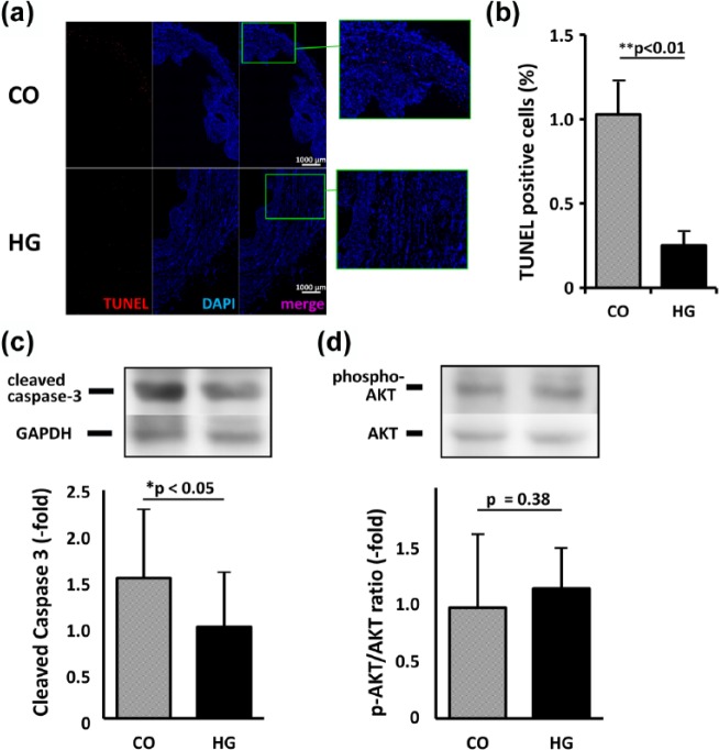
Figure 5.
Apoptosis in heart tissue. (a) The composite images of the heart stained with TUNEL (red) and DAPI (blue) were shown. When compared to the heart injected with cells only (CO), fewer TUNEL-positive cells were observed in the heart injected with cells in hydrogel (HG) (bar = 1000 µm). (b) The ratio of TUNEL-positive cells per nuclei (DAPI-positive) was 1.03% ± 0.56% versus 0.25% ± 0.19% for CO versus HG, respectively (p < 0.01). (c and d) Quantitative analysis of the bands in the Western blot revealed that the expression of cleaved caspase-3 was significantly higher in the CO group than in the HG group (1.54-fold, p < 0.05). (c) Although the ratio of phosphorylated AKT to total AKT tended to be higher in the HG heart, the difference was not statistically significant (1.17-fold, p = 0.38).

