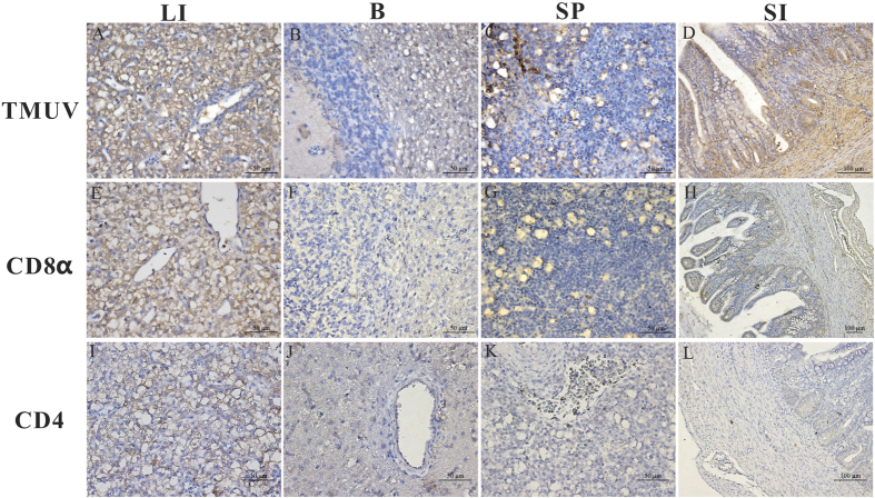
Figure 2. The location and density of TMUV antigen, CD4 and CD8α molecule in the liver (LI), brain (B), spleen (SP), and small intestine (SI).
Geese were humanly killed 5 days post infection by viruses. These protein locations in the different tissues of H9N2-infected birds were detected by IHC assay. Positive virus signals were detected, cells positive for CD4 or CD8α antigen appeared dark brown using immunohistochemical staining, and sections were counterstained with haematoxylin. Rabbit polyclonal antibody against TUMV E protein was prepared by our laboratory. The dilution folds of mouse anti-duck monoclonal CD4 antibodies and mouse anti-goose monoclonal CD8α antibodies were both 1:100. Incubation of goat anti-mouse or goat anti-rabbit secondary antibody was performed by the protocols of the immunoassay kit. Liver (A,E,I), brain (B,F,J), spleen (C,G,K) and small intestine (D,H,L).

