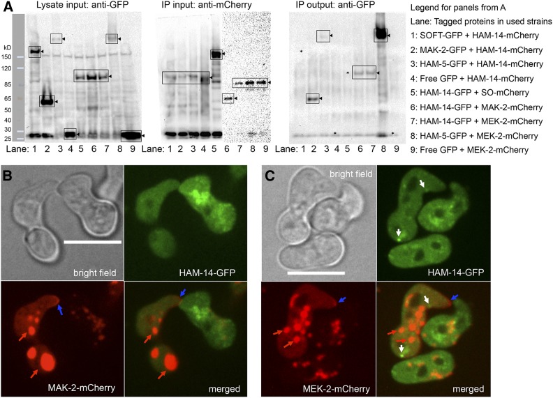
Figure 6.
Interaction of HAM-14–GFP with MAK-2–mCherry and MEK-2–mCherry. (A) Left shows Western blot of input protein samples from 5-hr-old germlings probed with anti-GFP antibodies (lane 1, SOFT-GFP; lane 2, MAK-2–GFP; lanes 3 and 8, HAM-5–GFP; lanes 4 and 9, free GFP; and lanes 5–7, HAM-14–GFP). Black triangles point to the bands of interest (boxed) and ladder on the left shows protein marker. Middle consists of two Western blots showing anti-mCherry immunoprecipitated input protein samples from 5-hr-old germlings probed with anti-mCherry antibodies (lanes 1–4, HAM-14–mCherry; lane 5, SOFT-mCherry; lane 6, MAK-2–mCherry; and lanes 7–9, MEK-2–mCherry; black triangles point to the bands of interest). Right is a Western blot of protein samples immunoprecipitated by anti-mCherry antibodies and subsequently probed by anti-GFP antibodies. Lane 2, MAK-2–GFP; lanes 3 and 8, HAM-5–GFP; lanes 6 and 7, HAM-14–GFP; black triangles point to the bands of interest. Lanes lacking co-immunopreciptation signal (1, 4, and 5) are marked with *. Legend on the right shows strains/lanes. (B) HAM-14–GFP and MAK-2–mCherry localization during germling fusion. Top is a bright field image (Bar, 10 µm); top right shows HAM-14–GFP fluorescence; bottom left shows MAK-2–mCherry fluorescence; and bottom right shows the composite of HAM-14–GFP and MAK-2–mCherry fluorescent images. Blue arrows point to MAK-2–mCherry crescent at the tip. Red arrows point to mCherry fluorescence visible in vacuoles, which has been observed previously in germlings (Jonkers et al. 2014). (C) Composite of HAM-14–GFP and MEK-2–mCherry localization during germling fusion. Top left is a bright field image (Bar, 10 µm); top right shows HAM-14–GFP fluorescence; bottom left shows MEK-2–mCherry fluorescence; and bottom right shows the composite of HAM-14–GFP and MEK-2–mCherry fluorescent images. White arrows point to HAM-14–GFP fluorescent puncta, blue arrows point to MEK-2–mCherry crescent at the tip, and red arrows point to mCherry fluorescence visible in vacuoles.

