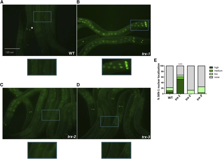
Figure 1.
TRX-1 negatively regulates nuclear localization of intestinal SKN-1. (A–D) Fluorescence microscopy was used to analyze the intestinal nuclear localization of SKN-1 (SKN-1B/C::GFP) upon the loss of trx-1, trx-2, or trx-3. Only upon the loss of trx-1 did SKN-1::GFP accumulate in intestinal nuclei. Asterisk in A depicts constitutive SKN-1B/C::GFP localization in the nucleus of the ASI neurons. Worms were visualized using a 20× objective. Blue boxes indicate the portion of the micrograph field that is magnified in the boxes below each micrograph. (E) Percentage of SKN-1::GFP nuclear localization was categorically scored and quantified as described in Materials and Methods. The percentage of SKN-1 nuclear localization increased threefold upon loss of trx-1 (P-value < 0.0001 as compared to wild type). Percentages are an average of three biological replicates (n = 100 worms per replicate).

