Figure 1.
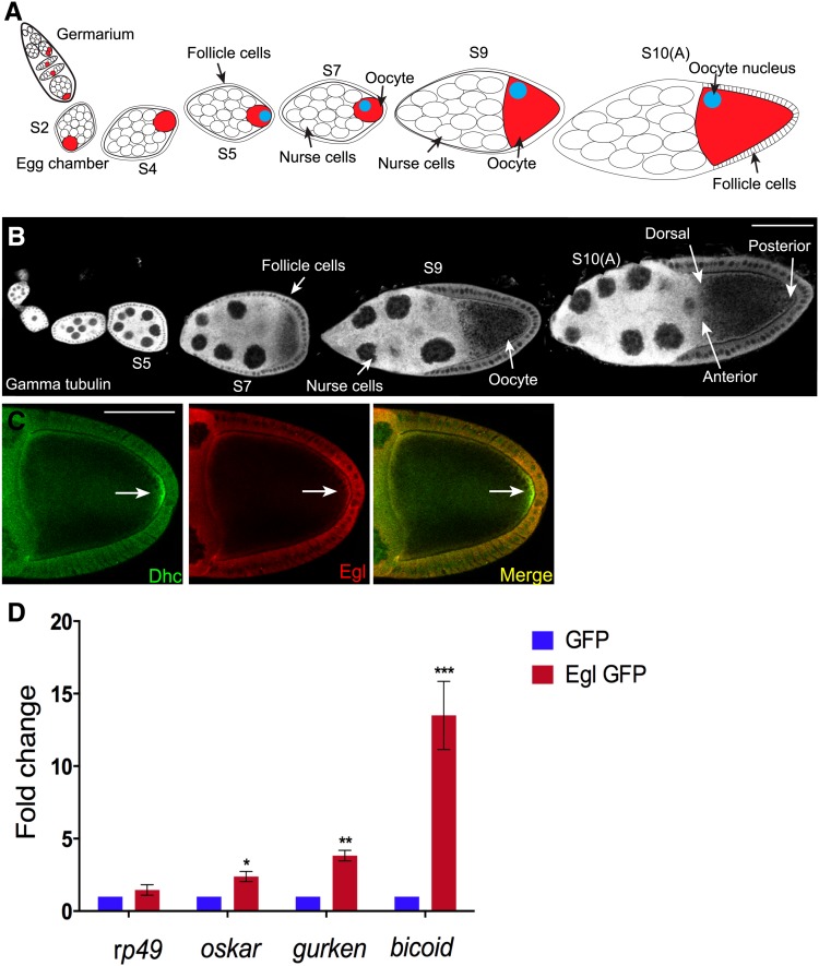
The localization and mRNA binding properties of Egl. (A) Schematic of a Drosophila ovariole. The germline stem cells and their niche reside at the anterior tip of the ovariole in a region known as the germarium. The stem cell divides to produce a daughter cell known as a cystoblast. The cystoblast undergoes four rounds of cell division with incomplete cytokinesis to produce a cyst containing 16 germ cells. One of these 16 germ cells will become the oocyte (red cell); the rest will assume nurse cell fate. During maturation within the germarium, the oocyte comes to reside at the posterior of the cyst. Also within the germarium, the cyst becomes surrounded by a layer of somatic cells known as follicle cells. This structure is now referred to as an egg chamber. The egg chamber progresses through 14 stages of morphogenesis before it is competent for fertilization. Egg chambers from the following stages are indicated in the schematic: stage 2 (S2), stage 4 (S4), stage 5 (S5), stage 7 (S7), stage 9 (S9), and stage 10A (S10A). Between stages 5 and 7, signaling events between the oocyte and the overlying follicle cells result in reorganization of oocyte microtubules. As a consequence, the oocyte nucleus migrates from the posterior to the dorsal–anterior margin (blue circle). (B) Representative Drosophila ovariole from a wild-type strain. The egg chambers were stained with an antibody against γ-tubulin. (C) Ovaries were dissected from wild-type flies and fixed and processed for immunofluorescence using antibodies against Dynein heavy chain (Dhc, green) and Egl (red). The arrow indicates enrichment of Dhc at the posterior pole. (D) Ovaries were dissected from flies expressing either GFP or Egl-GFP in the female germline. Lysates were prepared and incubated with GFP-Trap beads. The coprecipitating RNAs were reverse transcribed and processed for qPCR using primers against the indicated genes. The level of coprecipitating rp49, osk, bcd, and grk was normalized to the level of coprecipitating γ-tubulin mRNA. rp49 and γ-tubulin represent mRNAs that are not asymmetrically localized within the egg chamber. The entire experiment was done in triplicate. The error bars represent standard deviation. *P = 0.033, **P = 0.0013, and ***P = 0.0009, unpaired t-test. Bar, 50 μm.

