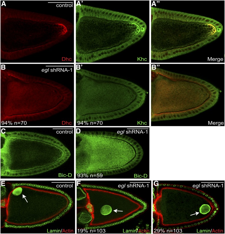
Figure 5.
Delocalization of Dynein, Kinesin, BicD, and the oocyte nucleus in Egl-depleted egg chambers. (A and B) Ovaries from flies expressing a control shRNA (A) or egl shRNA-1 (B) were fixed and processed for immunofluorescence using antibodies against Dhc (red) and Khc (A′ and B′, green). A merged image is also shown (A′′ and B′′). (C and D) Ovaries from flies expressing a control shRNA (C) or egl shRNA-1 (D) were fixed and processed for immunofluorescence using an antibody against BicD. (E–G) Ovaries from flies expressing a control shRNA (E) or egl shRNA-1 (F and G) were fixed and processed for immunofluorescence using an antibody against Lamin DmO (green). The egg chambers were also counterstained with Phalloidin (red). The arrow in E indicates the normal localization of the oocyte nucleus. The arrows in F and G indicate the mislocalized oocyte nucleus in Egl-depleted egg chambers. The number of egg chambers scored, as well as the penetrance of each phenotype, are indicated. Bar, 50 μm.

