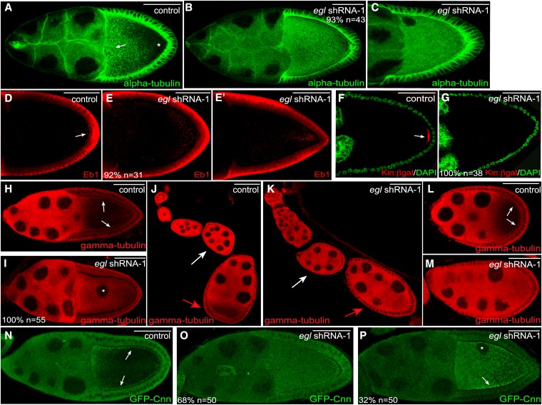
Figure 6.
Microtubule organization in Egl-depleted egg chambers. (A–C) Ovaries from flies expressing a control shRNA (A) or egl shRNA-1 (B and C) were fixed and processed for immunofluorescence using an antibody against α-tubulin. There is a high density of microtubules at the anterior margin of control oocytes (A, arrow). The posterior of control oocytes contains a much lower density of microtubules (A, asterisk). (D–E′) Ovaries from flies expressing a control shRNA (D) or egl shRNA-1 (E and E′) were fixed and processed for immunofluorescence using an antibody against Eb1. The arrow in D indicates the posterior enrichment of Eb1 foci in control egg chambers. (F and G) Ovaries from control flies expressing Kinesin β-gal (F, Kin:β-gal, red) or Kinesin β-gal along with egl shRNA-1 (G) were fixed and processed for immunofluorescence using an antibody against β-galactosidase. The egg chambers were also counterstained with DAPI (green). The arrow in F indicates the posterior localization of Kin:β-gal in control egg chambers. (H–M) Ovaries from flies expressing a control shRNA (H, J, and L) or egl shRNA-1 (I, K, and M) were fixed and processed for immunofluorescence using an antibody against γ-tubulin. The arrows in H indicate the cortical enrichment of γ-tubulin in control stage 10 egg chambers. The asterisk in I indicates the mislocalized oocyte nucleus. The white arrows in J and K indicate a stage 5 egg chamber and the red arrows in these panels indicate a stage 7 egg chamber. The arrows in L indicate the cortical enrichment of γ-tubulin in an early stage 9 egg chamber. (N–P) Ovaries from flies expressing GFP-Cnn (N) or from flies expressing GFP-Cnn and egl shRNA-1 (O and P) were fixed and processed for immunofluorescence using an antibody against GFP. Expression of the shRNA was under control of the early-stage maternal α-tubulin driver. Arrows in N indicate the cortical enrichment of GFP-Cnn in the control strain. The arrow in P indicates the residual cortical enrichment of GFP-Cnn in a subset of Egl-depleted egg chambers. The asterisk indicates the mislocalized oocyte nucleus. The number of egg chambers scored, as well as the penetrance of each phenotype, are indicated. Bar, 50 μm.

