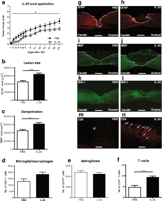
Fig. 1.

Local application of IL-25 decreases functional outcome and increases lesion size, demyelination, and T cell infiltration following SCI in mice. (a) Mice receiving local application of IL-25 show a statistically significant decrease in functional outcome when compared to those receiving PBS, as measured by the BMS (*p < 0.05), n = 9–10 mice/group. (b) Lesion size and (c) demyelinated area were quantified by staining for (g, h) GFAP and (i, j) MBP, respectively, as depicted by the dotted white line. Image analysis revealed a significant increase in (b) lesion size and (c) demyelinated area in animals treated locally with IL-25, compared with the PBS control group. Quantification of (d) Iba-1+ and (e) GFAP+ cells after SCI using TissueQuest software revealed no significant difference in (k, l) microglia/macrophages numbers or (g, h astrogliosis between animals receiving PBS or IL-25. (f) Significantly more CD4+ T cells are present in the spinal cord sections of the (n) IL-25-treated mice, compared with (m) PBS-treated mice, 3 weeks after SCI. Scale bars of representative photomicrographs: (g–l) = 500 μm, m + n = 50 μm. Data represent mean ± SEM. ***p < 0.001, **p < 0.01, n = 5–6 mice/group
