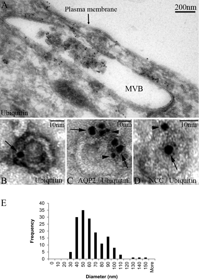
Fig. 2.
Identification of ubiquitin in human MVBs and urinary exosomes by immunogold electron microscopy. (A) Ubiquitin is readily detected in association with internal luminal vesicles of an MVB within a human kidney epithelial cell. Exosomes isolated from human urine labeled with ubiquitin (B) or ubiquitin plus AQP2 (C) or NCC (D); arrows indicate ubiquitin (5 nm gold particles) and arrowheads indicate AQP2 or NCC (2 nm gold particles). (E) Size distribution of low-density membrane vesicles from 200,000 × g pellet fraction (n = 159).

