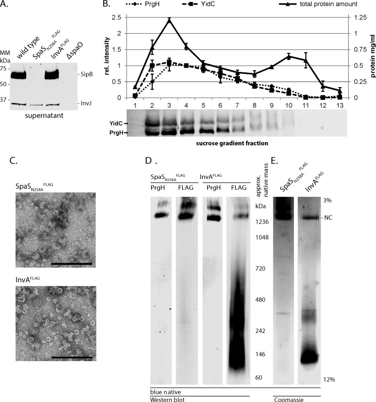
Fig. 3.
Purification of needle complexes by immunoprecipitation and blue native-PAGE. (A) Type III secretion into culture supernatant of wild-type and indicated mutant strains was profiled by immunodetection of the early substrate InvJ and the intermediate substrate SipB. ΔspaO denotes a negative control strain defective in type III secretion. (B) Membrane fractions containing needle complexes were identified by immunodetection of the external inner membrane ring protein PrgH. The inner membrane protein YidC served as a marker for the inner membrane. Quantification of the protein bands and of total protein content of the fractions was graphed. The average of three independent experiments is shown. Error bars show the standard deviation. (C) Electron micrographs of immunoprecipitated needle complexes, scale bar = 100 nm. (D, E) Blue native-PAGE of immunoprecipitated needle complexes; (D) shows immunodetection of the indicated proteins; (E) shows a Coomassie-stained gel of immunoprecipitated material.

