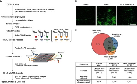
Fig. 1.
Comprehensive proteome profiling of retina tissues. A, overall scheme describing sample preparation, proteome profiling, and data analysis. See text for detailed description of each experimental step (“Experimental Procedures” and “Results”). B, VEGF-induced retinal vascular hyperpermeability. Intravitreally injected VEGF (100 ng)-induced vascular leakage of the superficial vascular plexus in the retina of C57BL/6 mice. Cotreatment of VEGF with anti-VEGF antibody (1 μg) effectively prevented the phenotype of increased permeability demonstrated by the leakage of FITC-dextran. Scale bar, 200 μm. C, comparison of our retinal proteome with two previously reported proteomes associated with VEGF-induced vascular permeability. The Venn diagram shows the relationships between the three retinal proteomes. The numbers of the detected proteins (see “Results” for the detection criteria) in the three studies and the genes encoding the proteins are shown in the table.

