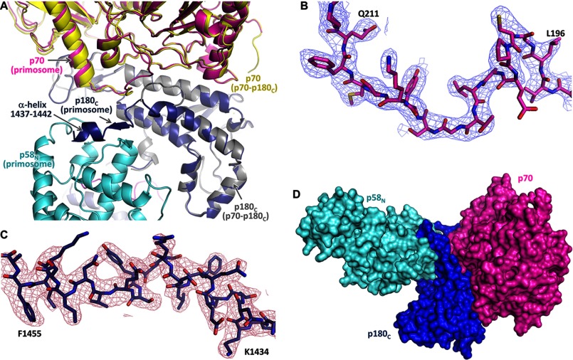
FIGURE 5.
The regions disordered in Polα are structured in primosome. A, superimposition of p180C-p70 (PDB code 4Y97) and primosome structures. In primosome, p180C and p70 are colored magenta and blue; in p180C-p70, they are colored yellow and gray. B and C, 2Fo − Fc Fourier map (contour level at 1σ) for p70 residues 195–212 and p180C residues 1433–1456, respectively. D, the relative position of p70 and p58N is stabilized by p180C.

