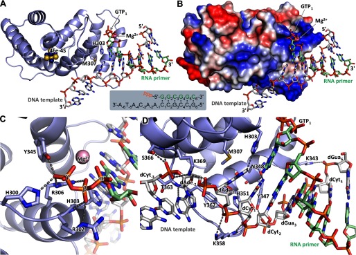
FIGURE 6.
Structure of p58C in complex with the RNA-primed DNA template. A, specific recognition of the template/primer junction at the 5′ terminus of the primer by p58C. B, the surface electrostatic potential of p58C shows docking of the template and the 5′-triphosphate of the primer on two separate positively charged areas (colored blue). C, hydrogen bonds (dashed lines) between p58C and the β- and γ-phosphates of GTP1. D, hydrogen bonds between p58C and the template.

