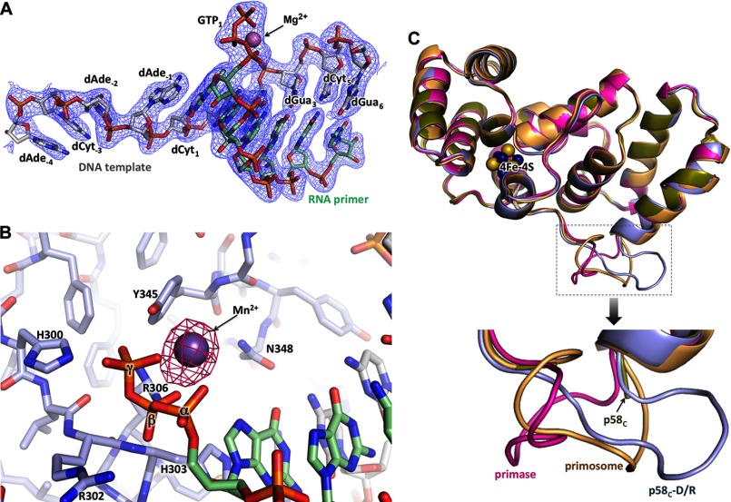
FIGURE 7.
Details of p58C interaction with a template/primer. A, electron density map for the DNA/RNA duplex and the triphosphate coordinating Mg2+. The carbons of DNA and RNA are colored green and gray, respectively. B, Fo − Fc Fourier map (contour level at 5σ) for Mn2+ coordinated by a 5′-triphosphate of RNA. C, alignment of p58C from different structures points to the flexibility of the DNA-interacting loop 354–366. The PDB accession numbers for the structures of the human primase and p58C are 4RR2 and 3Q36, respectively.

