FIGURE 1.
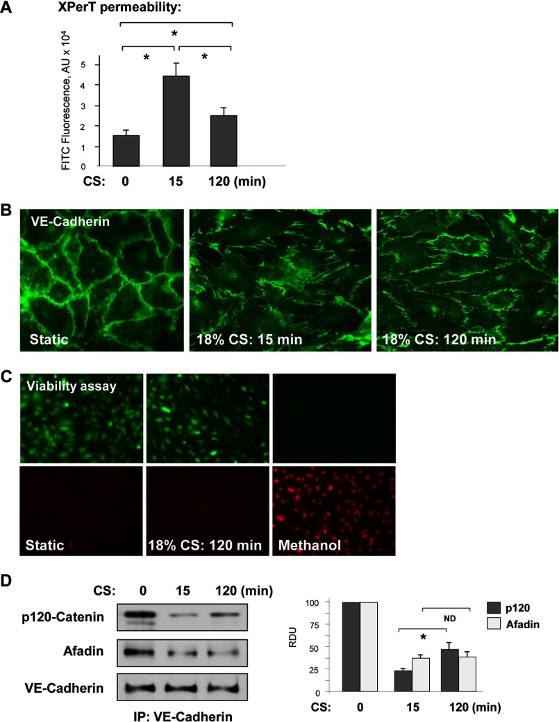
18% CS induced time-dependent changes in EC permeability, monolayer integrity, and adherens junction assembly. A, HPAEC grown in Biofex plates coated with biotinylated gelatin (0.25 mg/ml) were subjected to 18% CS for 15 or 120 min, followed by the addition of FITC-avidin (25 μg/ml, 3 min). Unbound FITC-avidin was removed, and FITC fluorescence was measured. n = 4; *, p < 0.05. B, adherens junction remodeling in control and stretched EC was examined by immunofluorescence staining for VE-cadherin. C, cell viability assay. HPAEC were subjected to 18% CS for 120 min or left under static conditions followed by staining with green fluorescent calcein-AM to indicate intracellular esterase activity and red fluorescent ethidium homodimer 1 to indicate loss of plasma membrane integrity. Methanol treatment (50%, 10 min) was used as a control. The results are representative of three independent experiments. D, VE-cadherin immunoprecipitation (IP) under nondenaturing conditions was performed from control EC or cells subjected to 18% CS. The presence of afadin and p120 catenin in immune complexes was examined by Western blot with corresponding antibody. The bar graph depicts quantitative densitometry analysis of Western blot data. n = 3; *, p < 0.05.

