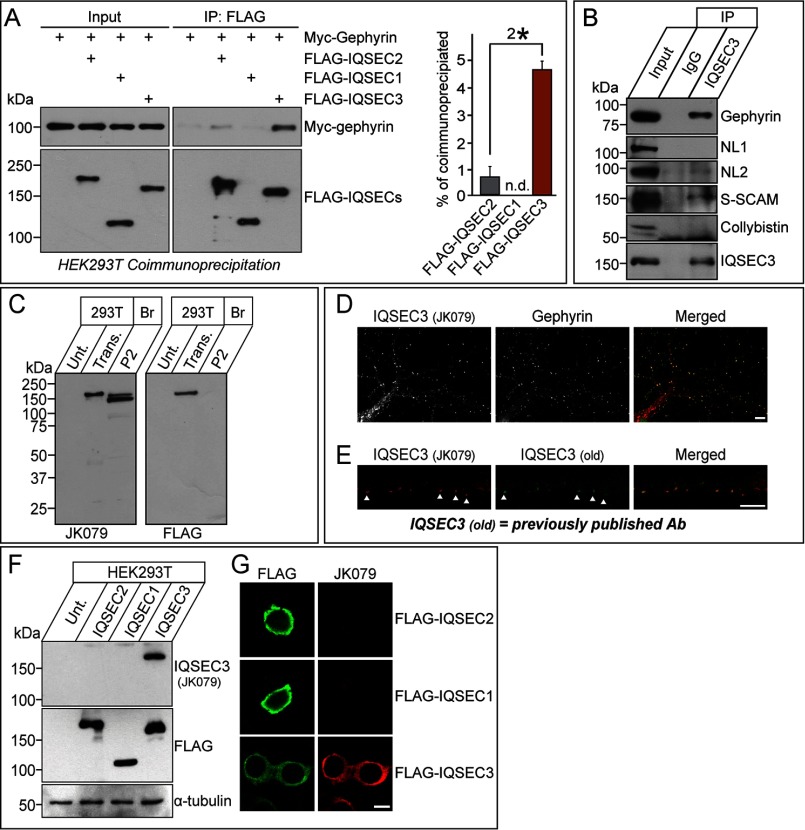
FIGURE 2.
Interaction of IQSEC3 with gephyrin in HEK293T cells, formation of an IQSEC3-gephyrin complex in rat brains, and characterization of IQSEC3 antibodies. A, co-immunoprecipitation (IP) experiment demonstrating that IQSEC2 and IQSEC3, but not IQSEC1, interact with gephyrin. HEK293T cells were transfected with FLAG-tagged IQSEC1 (FLAG-IQSEC1), FLAG-tagged IQSEC2 (FLAG-IQSEC2), or FLAG-tagged IQSEC3 (FLAG-IQSEC3) alone or together with Myc-tagged gephyrin (Myc-Gephyrin), and co-immunoprecipitation of IQSECs with gephyrin was assayed. A representative immunoblot visualized by ECL (left) and quantitative bar graphs (right) analyzing co-immunoprecipitation efficiency are shown. Note that maximum co-immunoprecipitation efficiency could not be achieved. Input, 5%. n.d., not determined. B, co-immunoprecipitation experiment in rat brains demonstrating that IQSEC3 forms complexes with gephyrin, NL-2, and S-SCAM but not with NL-1 or collybistin. Crude synaptosomal fractions of adult mouse brains were immunoprecipitated with anti-IQSEC3 antibody (JK079) and immunoblotted with the indicated antibodies. Equal amounts of rabbit IgG (IgG) were used as a negative control. Input, 5%. C, biochemical characterization of the anti-IQSEC3 antibody used in this study. Immunoblot analyses of the anti-IQSEC3 antibody, JK079, using rat brain crude synaptosomes (P2) and lysates from HEK293T cells transfected with a FLAG-tagged IQSEC3 expression vector (Trans.). The expression of FLAG-tagged IQSEC3 was confirmed by immunoblotting with an anti-FLAG antibody. Br., brain; P2, crude synaptosomes; Unt., untransfected HEK293T cell lysates. D, co-localization of IQSEC3 puncta with gephyrin puncta in rat cultured hippocampal neurons. Cultured neurons at DIV14 were immunostained with anti-IQSEC3 (JK079; red) and anti-gephyrin antibodies (green) and detected with Cy3- or FITC-conjugated secondary antibodies. Scale bar, 10 μm (applies to all images). E, validation of the in-house IQSEC3 antibody (JK079; red) by immunocytochemistry using DIV14 rat primary hippocampal cultured neurons. JK079-immunoreactive puncta co-localize with those obtained using previously published antibodies (old). Scale bar, 10 μm (applies to all images). F and G, cross-reactivity of the anti-IQSEC3 antibody. HEK293T cells, untransfected or transfected with the indicated IQSEC expression vectors, were immunoblotted or immunostained with anti-IQSEC3 (JK079) and anti-FLAG antibodies. JK079 is specific for IQSEC3. An anti-α-tubulin antibody was used for normalization. Scale bar, 10 μm (applies to all images).

