FIGURE 5.
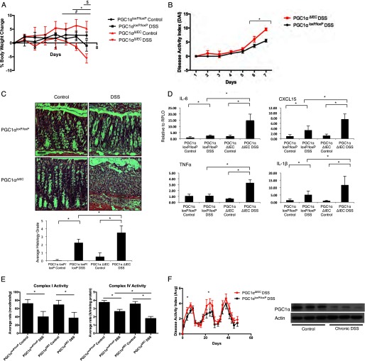
Intestinal epithelium-specific PGC1α knock-out mouse (PGC1αΔIEC) was subjected to 2% DSS for 7 days along with PGC1αloxP/loxP littermates (n = 8/group). When the % body weight was evaluated, significant differences were noted between PGC1αΔIEC control versus PGC1αΔIEC DSS-treated mice (#, p = ≤0.05), PGC1αΔIEC DSS versus PGC1αloxP/loxP DSS-treated mice (*, p ≤ 0.05), and PGC1αloxP/loxP control versus PGC1αloxP/loxP DSS-treated mice ($, p ≤ 0.05) (A). DAI score for PGC1αΔIEC mice was significantly higher than that for PGC1αloxP/loxP mice subjected to DSS colitis (B). Histologically, PGC1αΔIEC mice developed a more dramatic colitis, as quantified by average histology grade (C, scale bars, 100 μm). PGC1αΔIEC mice also demonstrated a significant increase in pro-inflammatory tissue cytokine release as compared with PGC1αloxP/loxP mice (D). Activity of the electron transport chain complexes I and IV were significantly decreased in both strains of mice after DSS exposure. However, complex IV activity was significantly decreased in PGC1αΔIEC mice subjected to DSS as compared with PGC1αloxP/loxP mice similarly treated (E). PGC1αΔIEC mice and PGC1αloxP/loxP mice were subjected to a chronic model of 2% DSS exposure (n = 16/group). Significant differences in DAI were noted in the first two cycles of DSS treatment but not the third cycle (F). As seen after acute DSS colitis, PGC1α is decreased in the intestines of PGC1αloxP/loxP mice after the completion of the chronic DSS model. *, p ≤ 0.05.

