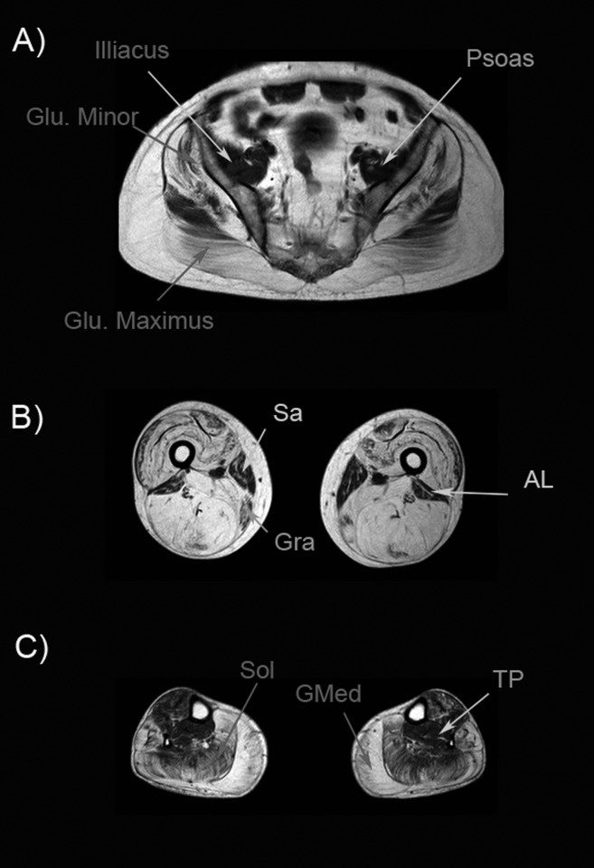
Figure 2.
Muscle MRI of a Becker muscle dystrophy patient. A) Pelvic image shows atrophy of glutei muscles (Gluteus minimus and maximus are shown in the image). Psoas and illiacus muscle are usually spared until late stages of the disease. B) Image of the thigh shows a complete atrophy of all muscles, except for sartorius (Sar), gracillis (Gra) and adductor longus (AL). C) Image of the legs showing a severe atrophy of gastrocnemius medialis (GMed) and soleus (Sol). In this patient, tibialis posterior (TP) was not involved.

