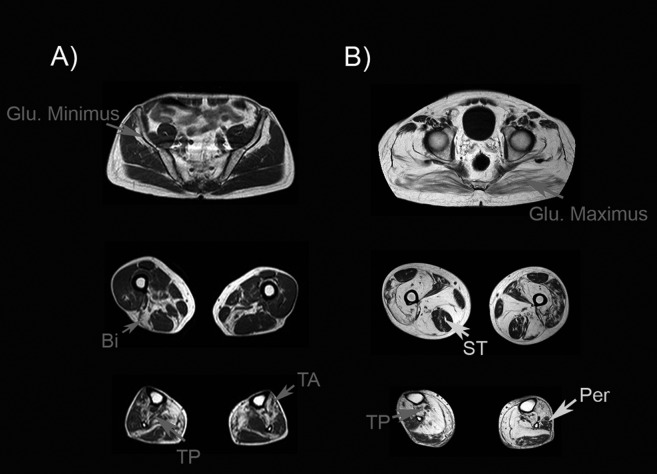
Figure 3.
Muscle MRI of two patients with mutations in the MYOT gene. A). Patient with mild weakness: muscle MRI shows mild involvement of gluteus minimus, biceps (Bi), tibialis anterior (TA) and tibialis posterior (TP). B) Patient with severe weakness: muscle MRI shows involvement of all glutei muscles, although gluteus maximus is less involved than gluteus minimus or medius. In the thighs, semitendinosus (ST) is not involved. In contrast, there is a clear involvement of the posterior muscles of the thighs. Tibialis posterior (TP) is more atrophic than peroneus muscle (Per).

