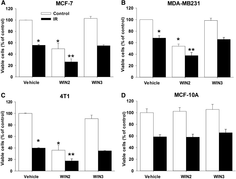
Fig. 2.
Enhanced antiproliferative effects of the combination of WIN2 and radiation. Cells were exposed to vehicle, WIN2, or WIN3 either alone or with 2 Gy radiation in MCF-7 (A), MDA-MB231 (B), 4T1 (8 Gy) (C), and MCF-10A (D) cells. Cells were treated with equieffective doses of WIN2 based on the concentration effect curves in Fig. 1 (12 µM for MCF-7 cells, 15 µM for MDA-MB231 cells, 30 µM for 4T1 cells, and 12 µM for MCF-10A cells). All experiments were analyzed for cell viability by trypan blue exclusion 96 hours after drug treatment (4T1 cells were analyzed 48 hours after treatment because of rapid growth rate). Data presented reflect the means of three or four individual experiments ± S.E.; *P < 0.05 versus vehicle and **P < 0.0156 compared with vehicle, drug treatment alone, and radiation alone. Two-way ANOVA reports: MCF-7 [F(2,12) = 12.8, P < 0.05]; MDA-MB231 [F(2,16) = 4.1, P < 0.05]; 4T1 [F(2,8) = 14.7, P < 0.01]; MCF-10A (P = 0.95).

