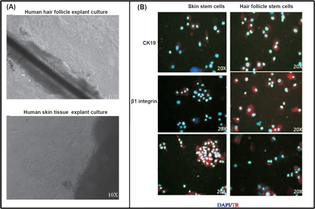
Fig. 1.
Expansion and charcaterization of skin and hair follicle stem cells. (A). Photomicrograph showing the explant culture of hair and skin tissue over fibronectin coated culture dish. The cells started coming out from the explant periphery after a week in culture media. (B). The cells were positive for the expression of CK-19, β1-integrin and CK-15 as revealed by immunofluorescence. DAPI (blue colour) was used as a nuclear stain. The secondary antibody was conjugated with Texas red (red colour).

