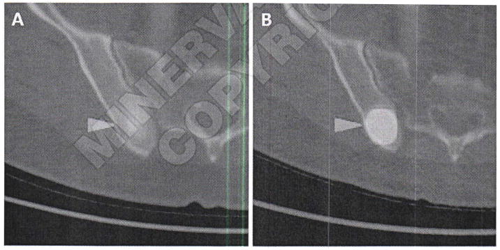
Figure 9.
Axial FDG PET/CT (A) and axial [18F]DCFPyL PET/CT (B) images of a patient with metastatic clear cell renal cell carcinoma involving the posterior aspect of the right iliac bone. Both images are set to the same quantitative scale, highlighting the increased tumor uptake achieved with [18F]DCFPyL in this case.

