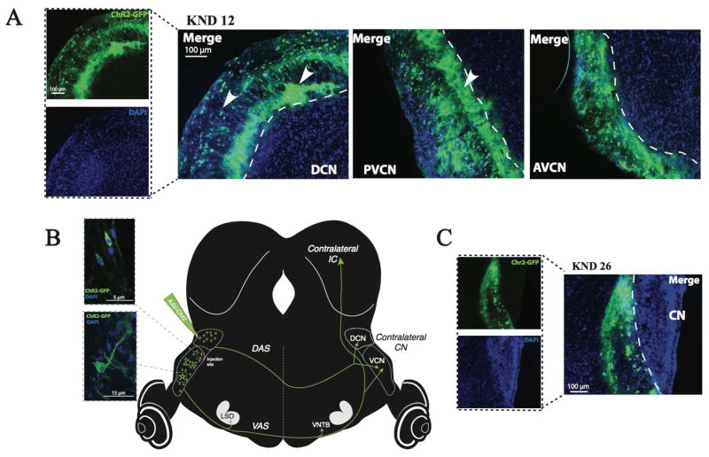
Figure 1.
Histological localization of ChR2-GFP. A) Fluorescent images of ChR2-GFP (top, left) and DAPI staining to indicate neurons (bottom, left) are merged into a composite view in the DCN. Other merged images indicate composites of staining in the PVCN and AVCN (all from case KND 12). Arrowheads indicate immunofluorescence label in fusiform cell layer and extracellular label in deeper DCN that obscures cell-type identification. B) High-magnification confocal images (merged DAPI and ChR2-GFP staining) of CN cell bodies. Schematic illustrates positions of ChR2 expression in cell bodies of the CN and in anterogradely labeled axons in the dorsal and ventral acoustic stria (DAS and VAS), the contralateral CN, and contralateral IC. C) Fluorescent images of a ChR2- case (e.g. KND 26) in which the expression pattern is observed outside the CN (just medial to it). In this and other ChR2- cases, no neuronal labeling of auditory brainstem structures was observed.

