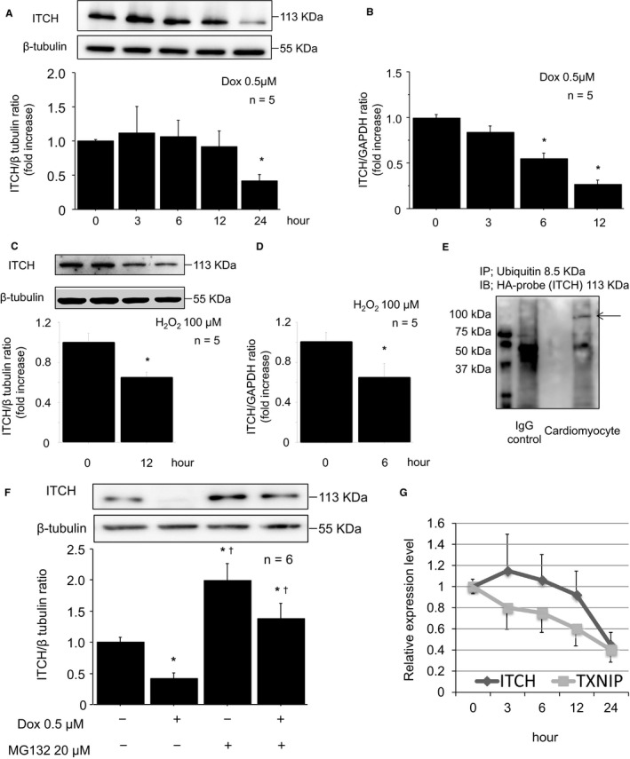
Figure 3.

Downregulation of ITCH was regulated by ubiquitin proteasomal degradation in reactive oxygen species–induced cardiotoxicity. A, Representative Western blot of ITCH in cardiomyocytes after Dox stimulation (0.5 μmol/L). B, mRNA levels of ITCH after Dox stimulation (0.5 μmol/L). C, Representative Western blot of ITCH in cardiomyocytes after H2O2 stimulation (100 μmol/L, 12 hours). D, mRNA levels of ITCH after H2O2 stimulation (100 μmol/L, 6 hours). Data are expressed as mean±SEM (n=5 per group, *P<0.05 vs 0 hour). E, Immunoprecipitation showed that ITCH bound with ubiquitin (arrowhead). HA‐ITCH–transfected cardiomyocyte lysates were immunoprecipitated with mouse anti–ubiquitin antibody and immunoblotted with rabbit anti–HA‐probe antibody. F, The ITCH decrease was reversed by pretreatment of MG132 (20 μmol/L, 2 hours). Data are expressed as mean±SEM (n=6 per group, *P<0.05 vs control, † P<0.05 vs Dox stimulation without MG132). G, Relative expression levels of TXNIP and ITCH after Dox stimulation (0.5 μmol/L). Dox indicates doxorubicin; HA, pRK5‐hem agglutinin; H2O2, hydrogen peroxide; IgG, immunoglobulin G; TXNIP, thioredoxin‐interacting protein.
