Figure 2.

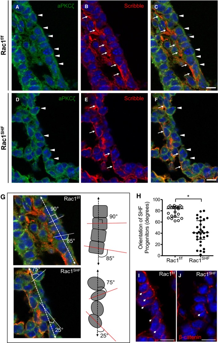
Disrupted apicobasal cell polarity and orientation in Rac1 SHF splanchnic mesoderm. A and B, In embryonic day 9.5 (E9.5) Rac1 f/f SHF progenitors, the basolateral domain is marked by Scribble (B, arrows), and the apical domain is marked by aPKCζ (A, arrowheads). The SHF progenitors have a distinct cuboidal shape, forming an organized epithelial layer (C). Polarity is disrupted in Rac1 SHF SHF progenitors in which the Scribble‐positive basal domains (E, arrows) and aPKCζ‐positive apical domains (D, arrowheads) of individual cells are no longer aligned with neighboring cells due to the rounded morphology of the SHF progenitors (F). C, Overlay of panels A and B. F, Overlay of panels D and E. The angle of the E9.5 SHF progenitor cell long axis (dashed line) was measured relative to the axis of the dorsal pericardial wall (dashed arrow line) to obtain the degree of orientation (G). Images in panel G are from panels C and F with an overlay of schematic axis and angle measurements. H, The angle of each SHF progenitor cell in the splanchnic mesoderm was measured and plotted. *P<0.05 by Mann–Whitney test, n=4 embryos per group. Each data point represents 1 SHF progenitor cell. Active (nonphosphorylated) β‐catenin marked cell–cell junctions in E9.5 Rac1 f/f SHF progenitors (I, arrows). In comparison, cell–cell junctions were disrupted in E9.5 Rac1 SHF SHF progenitors (J). Scale bars: 10 μm (A through F, I and J). SHF indicates second heart field.
