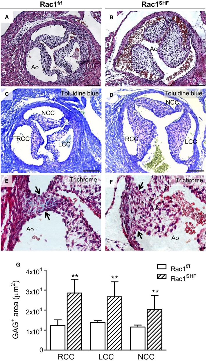
Figure 8.

Aortic valve defects in P0 Rac1 SHF hearts. More than 30% (9 of 28) of P0 Rac1 SHF hearts exhibited thick aortic valve leaflets (B) compared with the thin, remodeled valves of controls (A). Toluidine blue staining showed that GAG (light purple color) occupies the acellular space of Rac1 SHF and littermate valve leaflets (C and D). Masson's trichrome staining in Rac1 f/f aortic valves showed collagen in the commissure of valve leaflets (E, arrows), which was absent in Rac1 SHF aortic valves (F, arrows). The GAG‐positive area (light purple color) in each valve leaflet (C and D) was quantified in (G). **P<0.01 by Mann–Whitney test, n=5 to 6 hearts per group. Scale bars: 100 μm (A through D), 10 μm (E and F). Ao indicates aorta; GAG, glycosaminoglycans; LCC, left coronary cusp; NCC, noncoronary cusp; RCC, right coronary cusp; SHF, second heart field.
