
Figure 8.
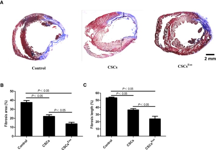
Histological analysis for myocardial infarction (MI) sizes in each group. Cardiac stem cells (CSCs)Exo were preconditioned with 400 μg/mL mesenchymal stem cells (MSCs) for 24 hours, and CSCs were injected into the peri‐infarct zones after MI induction. Heart samples were harvested 28 days after cell injection. A, Heart sections were stained with Masson trichrome: myocardium (red), scarred fibrosis (blue). The percentage of fibrotic area (B) and fibrosis length (C) was calculated and averaged (n=5) by using Image J software.