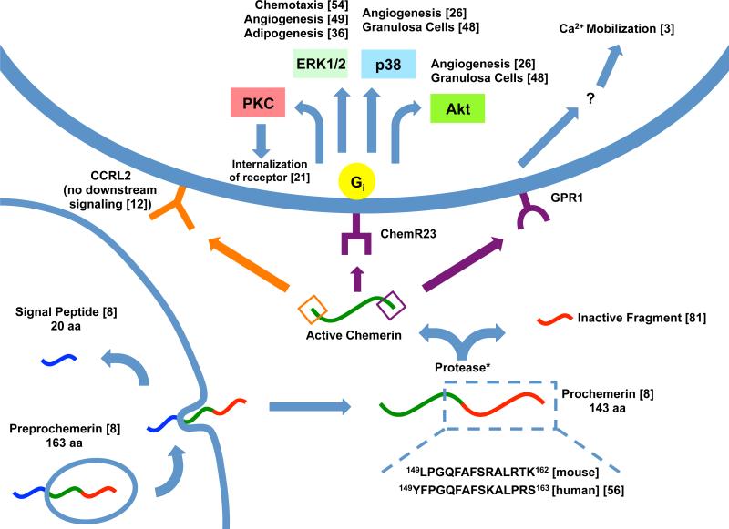
Figure 1. Processing and pathways of chemerin.
Chemerin is depicted with the N-terminus on the left and C-terminus on the right. When preprochemerin is released, the membrane-bound signal peptide (blue) is released and the protein becomes prochemerin [8]. This is then cleaved at various locations by a protease (*see Table 1), creating an inactive prochemerin fragment (red) and active chemerin (green) [81]. The N-terminus (orange) can bind CCRL2 [12] or the C-terminus (purple) can bind ChemR23 or GPR1. CHO-K1 cultured cells demonstrate Gi signaling to various pathways [8] which differ depending on the process being carried out. GPR1 has been shown to bind chemerin and induce Ca2+ mobilization but the specific mechanisms are still unknown [3].

